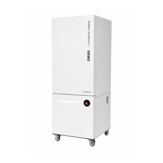
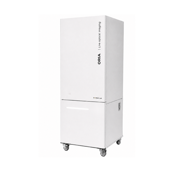
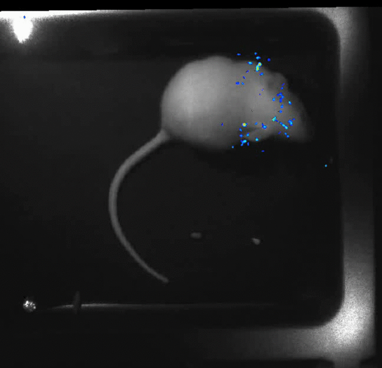
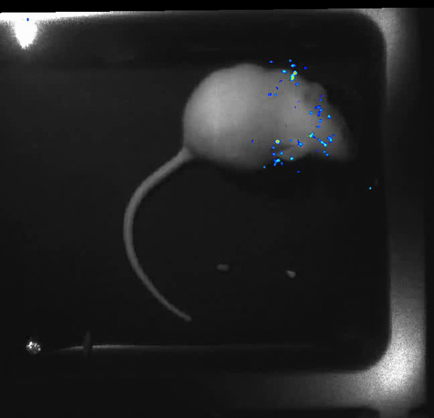
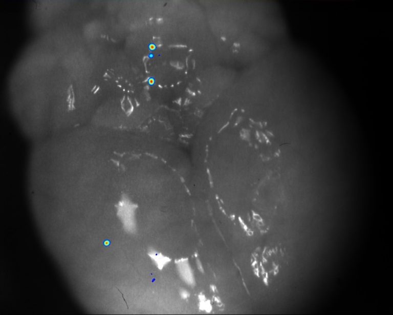
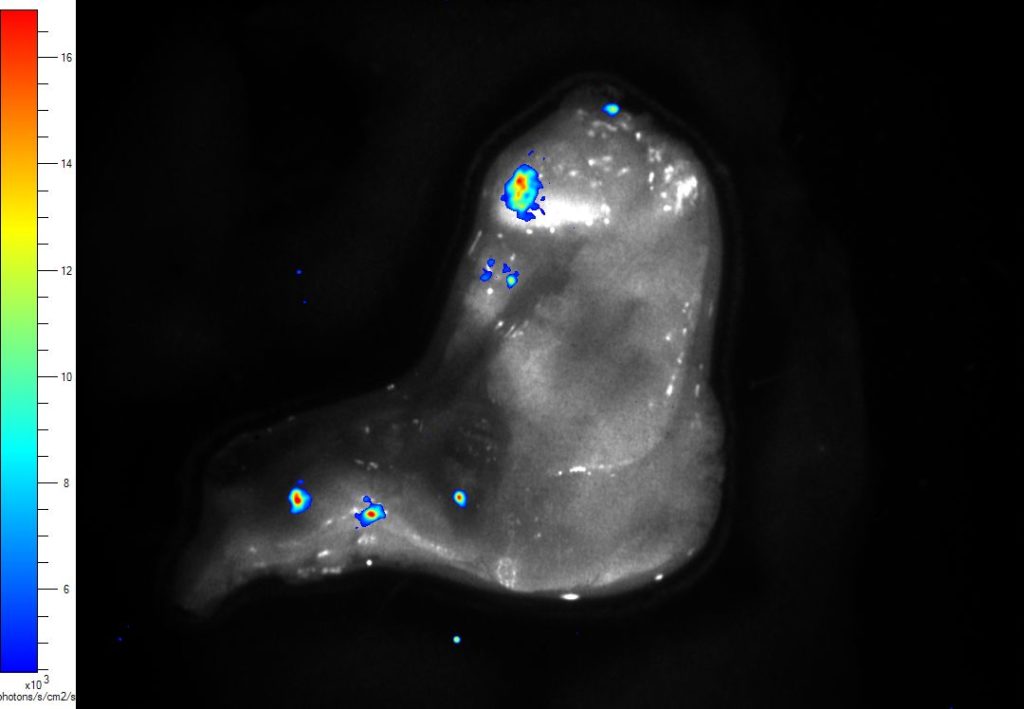
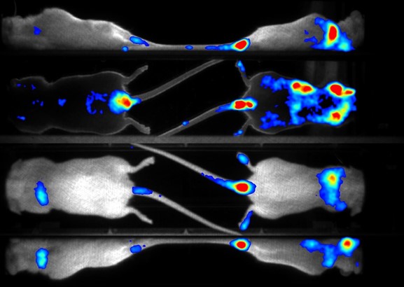
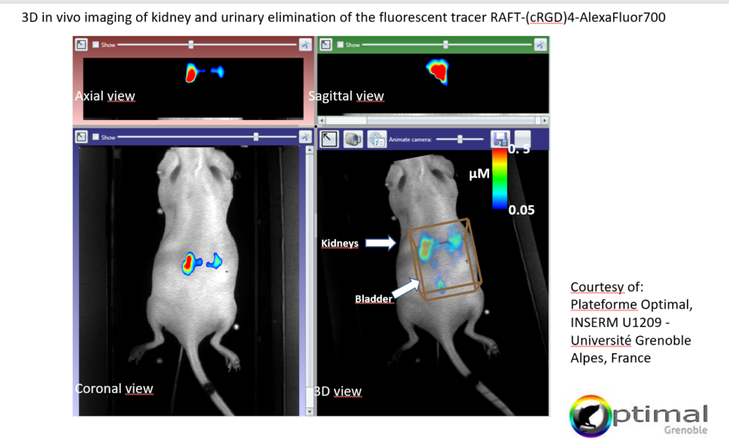
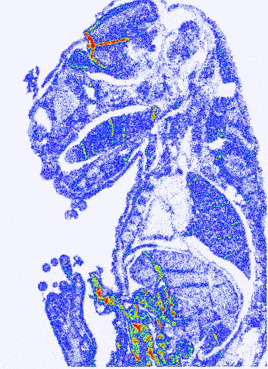
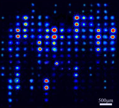
This technology makes it possible to count each photon individually emitted by the signal center. This method of quantification is called photon counting and leads to an almost perfect linearity as it sums each photon detected.
The photon counting technology combined with the high dynamic range of the optical chain allows the system to reach a global linear range over 106 and a linear range within a same image over 104.
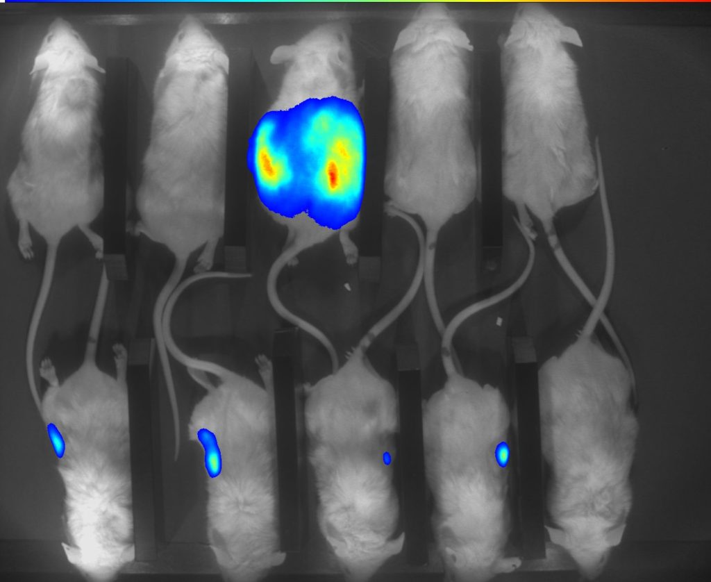
The high sensitivity opens the way towards bioluminescence detection to a few cells in subcutaneous tissues and a few hundred cells in deep tissues (livers, lungs, pancreas…).
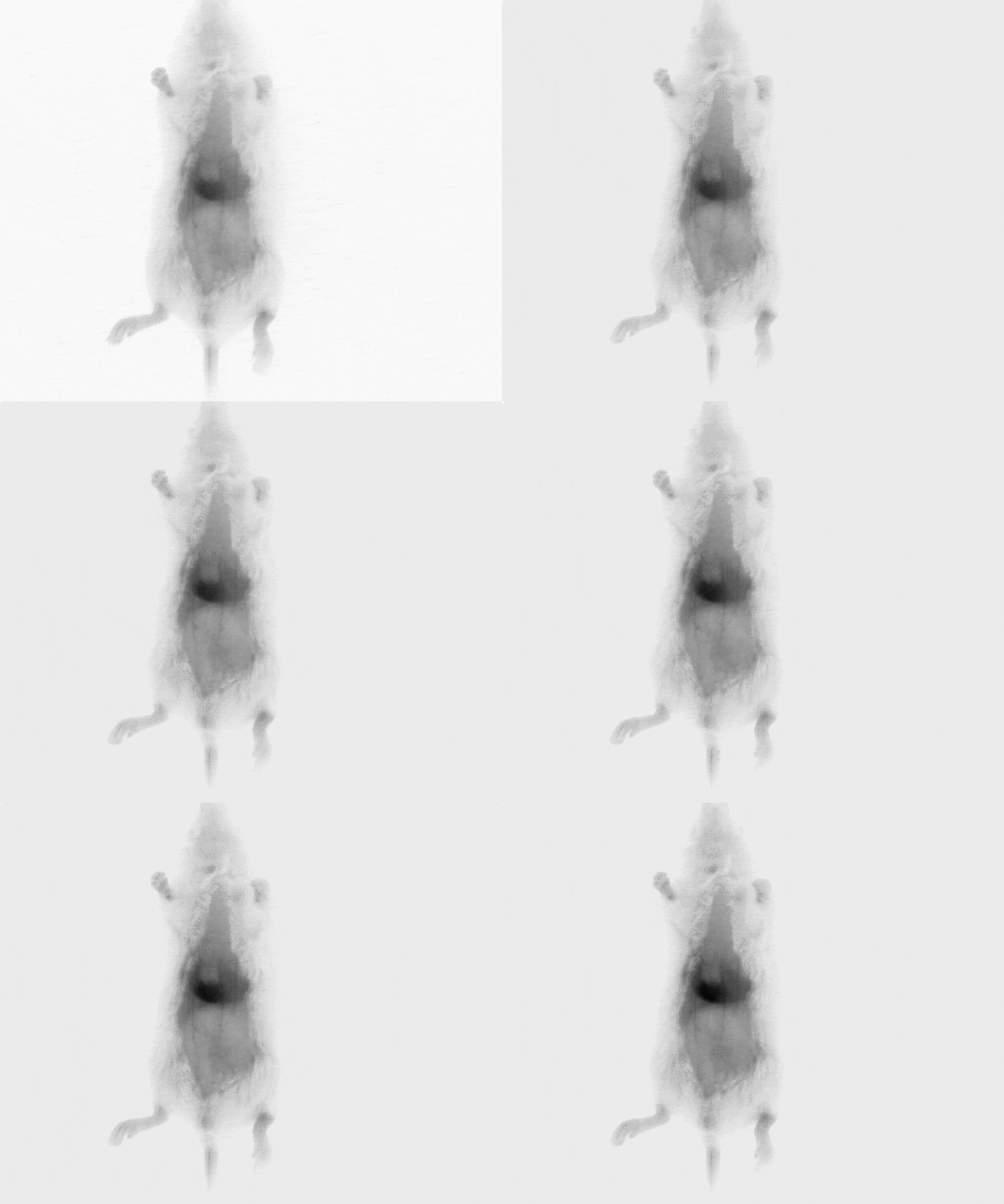
Detect the tiniest biological processes thanks to the unmatched sensitivity of our unique dynamic acquisition technology.
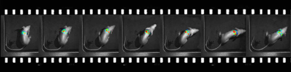
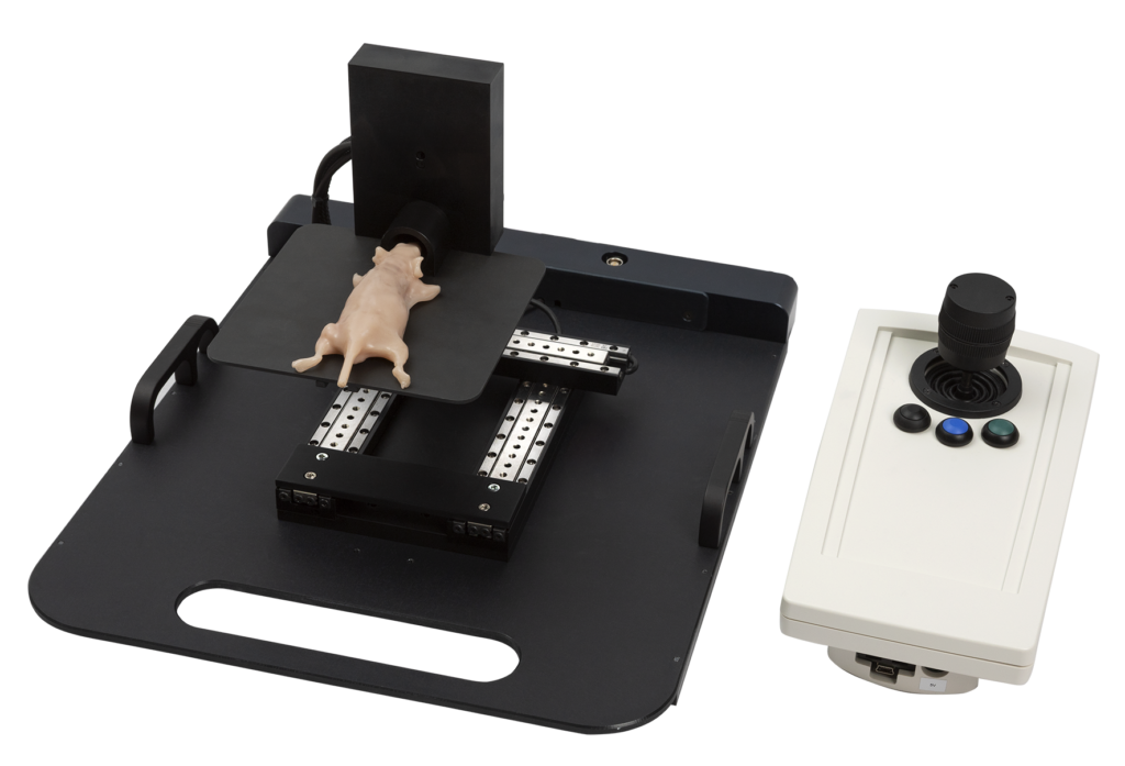
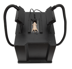
With the real-time technology of the ORSO system, you can even perform experiments with non-anesthetized, freely moving animals, opening previously unimagined new opportunities for research.
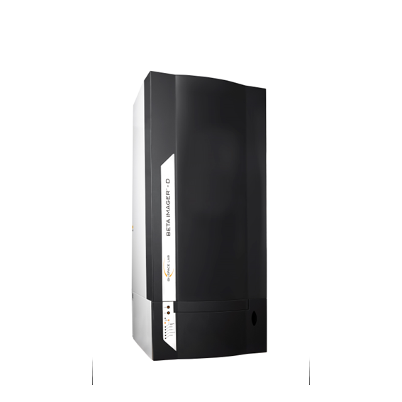
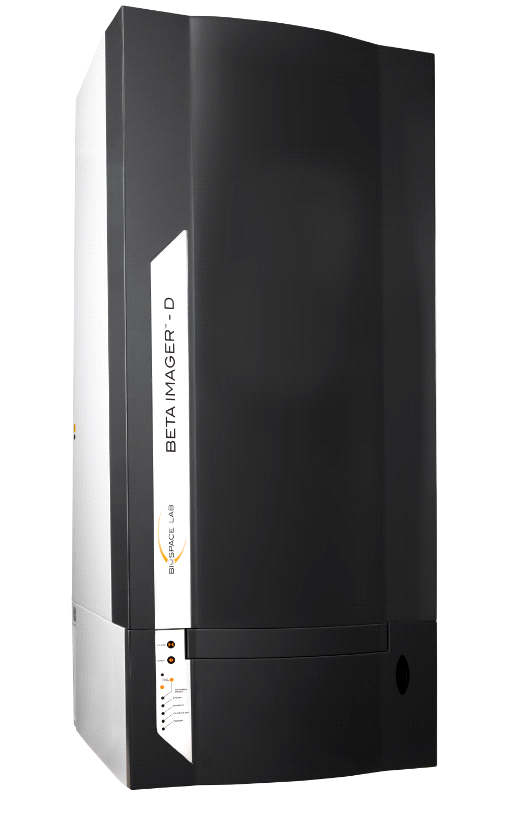
The BetaIMAGER™ tRACER is the system of choice for applications requiring the highest sensitivity and either high throughput, or larger sample sizes. With the BetaIMAGER tRACER, autoradiography becomes a fast and easy routine.
The BetaIMAGER™ tRACER is the system of choice for applications requiring the highest sensitivity and either high throughput, or larger sample sizes. With the BetaIMAGER tRACER, autoradiography becomes a fast and easy routine.
Tritium is a reliable beta emitter for measurement of biological samples. The big problem is that detection is slow. Traditional film and phosphor imaging methods can take weeks to complete. But now, there’s a better way to meet the high throughput needs of your lab: The BetaIMAGER tRACER system cuts your tritium imaging time from weeks to hours. Exceptional sensitivity can be obtained even with ultra-low energy tritium emissions: the tRACER system detects tritium levels as low as 0.007 cpm/mm2.
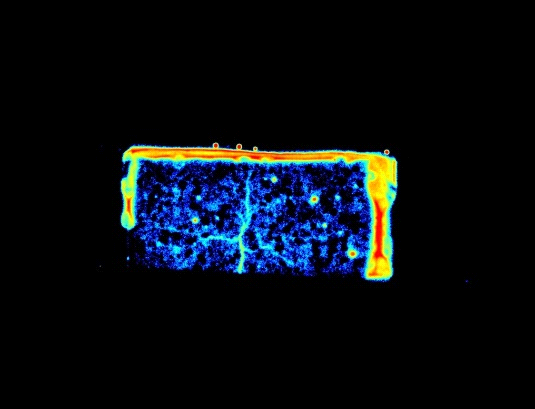
Tritiated sample. 18 h acquisition. Minimal visual range.
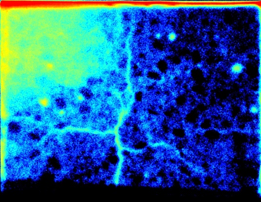
Tritiated sample. 5 h acquisition. Wider visual range than the sample
Sample activity was calculated using the acquisition shown in the figure on the left (exhibits the entire sample): 7.5 x 10^3 hpm (hits per minute) which translates to 6.82 nCi.
The ONLY high-resolution digital autoradiography solution.
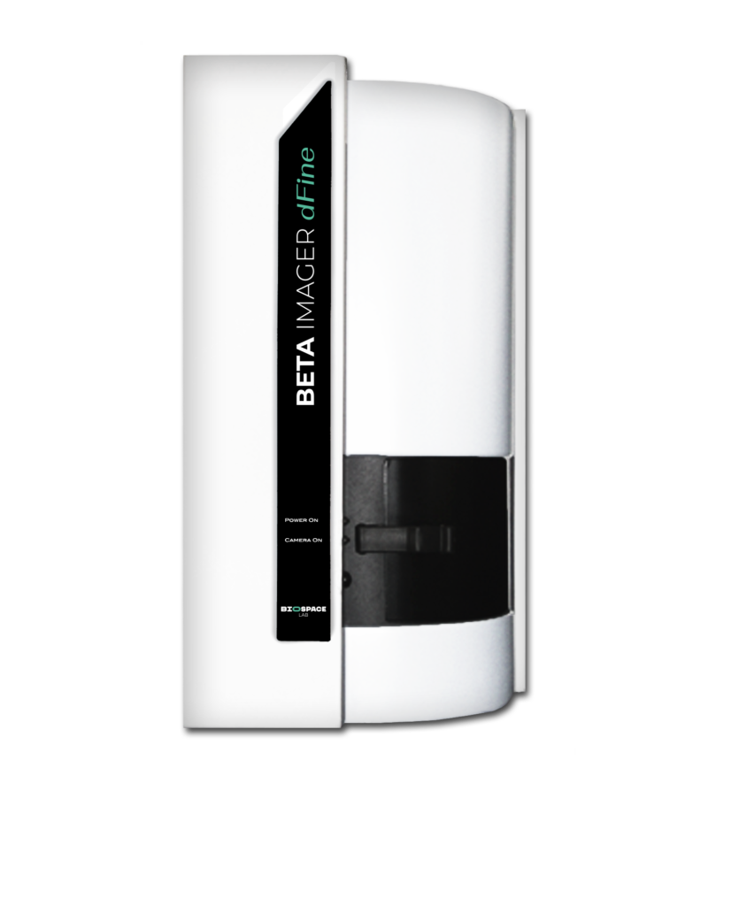
The BetaIMAGER™ DFINE is the system of choice for applications involving smaller tissue sections and where the best possible spatial resolution is required. Four samples, up to 24 x 32 mm each in size, can be loaded onto the automated stage for sequential image acquisition. Spatial resolution as high as 10 μm can be achieved with tritium. The DFINE is the perfect ex-vivo complement to PET and SPECT molecular imaging.
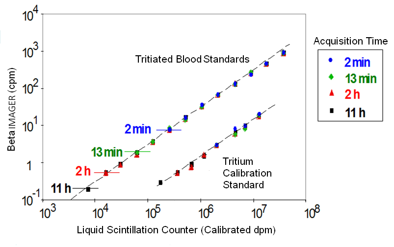
DETECT AND QUANTIFY HIGH AND LOW SAMPLE CONCENTRATIONS IN A SINGLE ACQUISITION
Linear dynamic range of the BetaIMAGER
Courtesy of Dr Alain Schweizer, Novartis
The BetaIMAGER systems count all emitted particles, regardless of their energy. This direct beta counting technology of the BetaIMAGER systems provides unequalled performance: the BetaIMAGER tRACER system is approximately 500X more sensitive and the BetaIMAGER dFINE system about 50X more sensitive to tritium than either X-ray film or storage phosphor screen. The BetaIMAGER tRACER system is unique in its ability to detect tritium levels as low as 0.007 cpm/mm2.
The BetaIMAGER systems provide precise localization and quantification of beta-emitting radioligands. The advanced image analysis tools of the M3Vision software allow for direct analysis of digital data and a better than 1:10,000 linear dynamic range ensuring accurate quantification over the widest possible range.
You can detect and quantify both high and low sample concentrations in a single data acquisition.
Biospace Lab developed an exclusive technology for the simultaneous detection and discrimination of multiple radioisotopes.
A single sample can be simultaneously labeled with several radioisotopes and the specific contribution from each label can be detected during a single data acquisition. Discrimination can be based on emission energy (2 radioisotopes), or on radioactive half-life (2 isotopes or more). Examples of dual-detection radioisotope pairs include 3H/14C, 3H/35S, 3H/32P or 33P and an example of a multi-label combination would be 99mTc/ 111In/18F.
This unique radioisotope discrimination feature allows the development of more rapid and accurate protocols for recording the distribution of several molecules simultaneously in a single specimen.
You can determine co-localization of different receptors, or receptors and ligands; Map complex functional activity with pathology; or Compare specific gene expression with a reference gene.
SAVE hours or days by acquiring images in real time thanks to our real time detection technology.
The exclusive real-time detection technology used in both systems displays and accumulates counts in real-time:
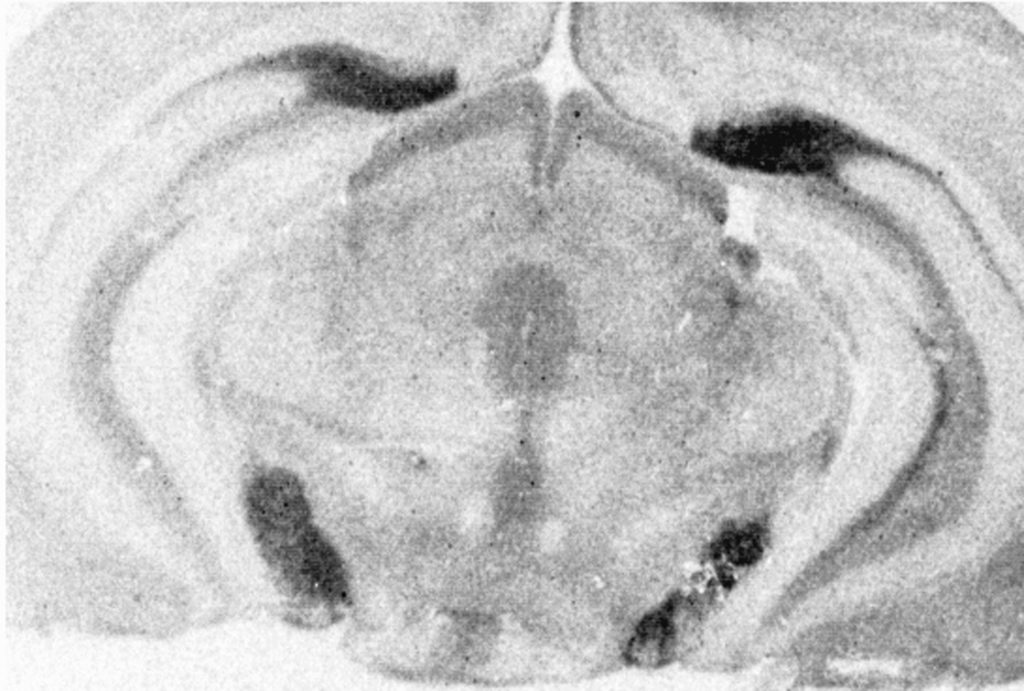
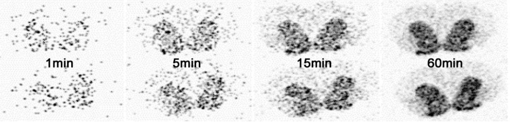
Real-time autoradiography: distribution of D1 receptors in rat brain striatum following tritiated ligand binding
X-Ray film or storage phosphor screen-based autoradiography requires a two-step blind acquisition process: expose and then scan
The real-time acquisition allows the instantaneous display of the image recorded by the camera and to follow the progression of the reconstitution of the final image. The image display is refreshed every 30 seconds allowing the user to evaluate it during the acquisition.
The unmatched sensitivity reduces your data acquisition time from weeks to hours.
You can see data accumulating in real-time and obtain the first results in minutes.
UNIQUE SYSTEM DETECTS AND QUANTIFIES BOTH HIGH AND LOW SAMPLE CONCENTRATIONS IN A SINGLE DATA ACQUISITION WITH BOTH HIGH SENSITIVITY AND RESOLUTION.
The BetaIMAGER™ systems count all emitted particles, regardless of their energy. This direct beta counting technology of the BetaIMAGER systems provides unequalled performance:
the BetaIMAGER tRACER system is approximately 500X more sensitive and the BetaIMAGER dFINE system about 50X more sensitive to tritium than either X-ray film or Storage Phosphor Screen. The BetaIMAGER™ tRACER system is unique in its ability to detect tritium levels as low as 0.007 cpm/mm2.
The BetaIMAGER systems provide precise localization and quantification of beta emitting radioligands. The advanced image analysis tools of the M3 vision software allow for direct analysis of digital data and a better than 1:10,000 linear dynamic range ensuring accurate quantification over the widest possible range.
Linear dynamic range of the BetaIMAGER
Courtesy of Dr Alain Schweizer, Novartis
You can detect and quantify both high and low sample concentrations in a single data acquisition.
Biospace Lab developed an exclusive technology for the simultaneous detection and discrimination of multiple radioisotopes
A single sample can be simultaneously labeled with several radioisotopes and the specific contribution from each label can be detected during a single data acquisition. Discrimination can be based on emission energy (2 radioisotopes), or on radioactive half-life (2 isotopes or more). Examples of dual-detection radioisotope pairs include 3H/14C,3H/35S, 3H/32P or 33P and an example of a multi-label combination would be 99mTc/ 111In/18F
This unique radioisotope discrimination feature allows the development of more rapid and accurate protocols for recording the distribution of several molecules simultaneously in a single specimen.
You can determine co-localization of different receptors, or receptors and ligands; Map complex functional activity with pathology; or Compare specific gene expression with a reference gene.
NO LOST DATA OR EXPERIMENTS CAUSED BY SATURATION.
Disintegrations are detected in real-time and the data display is continuously updated during acquisition. Under or overexposure of the sample are eliminated.
Longer acquisitions deliver higher image dynamics. The absence of limit on the duration of the acquisition allows the detection of weak signals and the improvement of resolution.
Use minimal amounts of radiolabeled compounds, reduce radioactive waste and minimize experimental costs as you can detect ultra-low level of bound radiolabeled ligand.
Thanks to their high sensitivity, BetaIMAGER systems can detect ultra-low levels of radiolabeled ligands in the sample. You can use minimal amounts of radiolabeled compounds and reduce radioactive waste as well as minimize your experimental costs.
Reduce the number of experiments thanks to the reproducibility of the system.
Non-destructive imaging and measurement: specimens are never damaged or destroyed during the process.
软件
The IMAGER systems are driven by the PhotoAcquisition and BetaAcquisition software application, which seamlessly control the entire system and its modules, and manage the imaging experiments.
Simple and user-friendly acquisition software.
Simplicity and user-friendly Acquisition software
Immediate visualization from the beginning of acquisition is one of the hallmarks making real-time imaging so practical. You can see the first signals after just a few seconds and can quickly stop the acquisition or extend it knowing that the signal will continue to accumulate until the desired endpoint.
Real-time feedback allows for optimized and automated control of some acquisition parameters such as the opening of the lens during the acquisition according to signal progression. These automatic time optimization capabilities simplify the use without blind configuration of the lens opening, integration time or binning level which must often be determined experimentally on the cooled CCD camera systems.
Simplicity of the analysis software
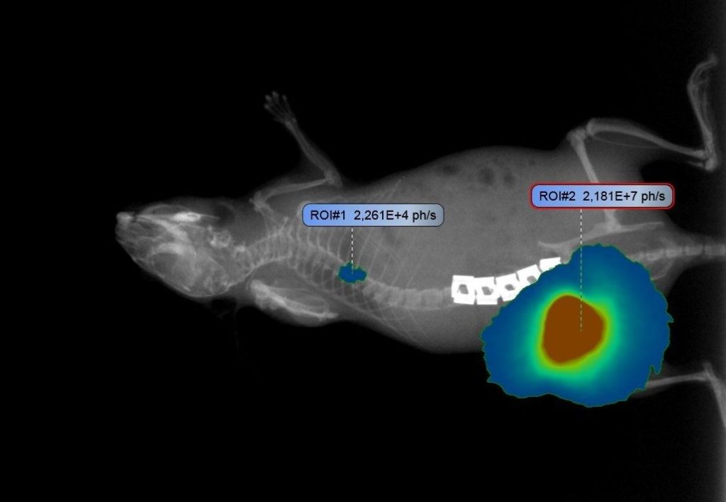
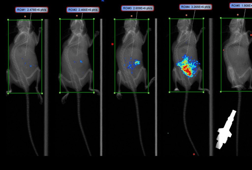
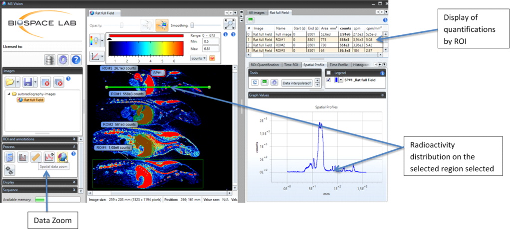
M3Vision analysis software allows users to visualize 2D and 3D images, freely adjust the signal display threshold, and perform quantifications based on desired ROIs such as geometrical forms or signal intensity thresholds.
ROI measurements provide total, maximum, minimum and average signal intensity values.
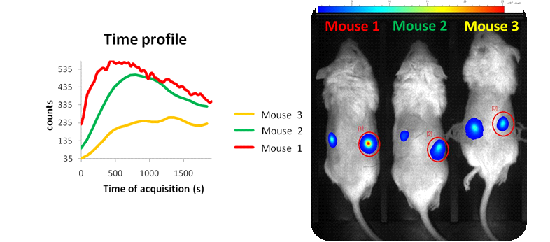
M3Vision makes time analysis possible, allowing quantifications at multiple ROIs at the plateau time. For high speed studies, an automatic selection tool of the temporal window to the signal plateau enables rapid analysis of each ROI.
Export all data, measurements and time profiles to Excel in a single click! For the longitudinal studies, all acquisitions linked to a study can be exported in a single Excel workbook that includes one tab per acquisition as well as a summary table of all the quantifications.
Software analysis tools include numerous functions to allow precise and automatic quantification:
The magic wand tool allows you to automatically draw ROIs determined by signal intensity thresholds. These ROIs can then be created and analyzed automatically by the software in a fraction of a second.
Automatic temporal analysis enables automated, streamlined and rapid quantification of each ROI in just a few seconds.
Traceability
When an acquisition is saved, ALL acquisition settings (including lens opening, plate position, acquisition time, grid speed, date and time) are also saved. User-generated comments can also be saved such as observations about the animals being imaged, and notable events that occurred before or during acquisition. This information is linked to the acquisition folder and is accessible through the analysis software.