Detect, quantify and monitor biological processes thanks to unmatched sensitivity and the benefits of kinetics recording.
The non-invasive nature of optical imaging allows the high throughput monitoring of your research interest, such as disease progression or infection course within the same group of animals. Several subgroups of animals can be analyzed in the context of one experiment, accelerating the studies and, thereby, reducing the number of animals needed to generate statistically significant data. In addition, by avoiding the need for invasive sampling procedures, optical imaging can also result in a reduction in the levels of stress and discomfort experienced by animals.
The PhotonIMAGER systems enable the in vivo acquisition of bioluminescence and fluorescence signals in small animals within the visible spectrum, ranging from violet to near infrared.
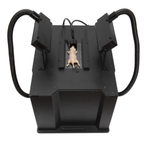
This system combines high sensitivity, very wide quantification dynamic range and high speed, with the capacity to image up to 10 animals per acquisition.
1- Detect the tiniest biological processes thanks to the unmatched sensitivity of our unique dynamic acquisition technology.
A single photon detected on the entry face of the intensified camera will generate an intense light spot easily detectable and recognizable by the CCD when intensified.
This technology allows to count individually each photon emitted by the signal center. This method of quantification is called photo counting and leads to an almost perfect linearity as it sums each photon detected.
The photo-counting technology combined with the high-dynamic of the optaical chain allow to reach a global linear range over 106 and a linear range within a same image over 104.
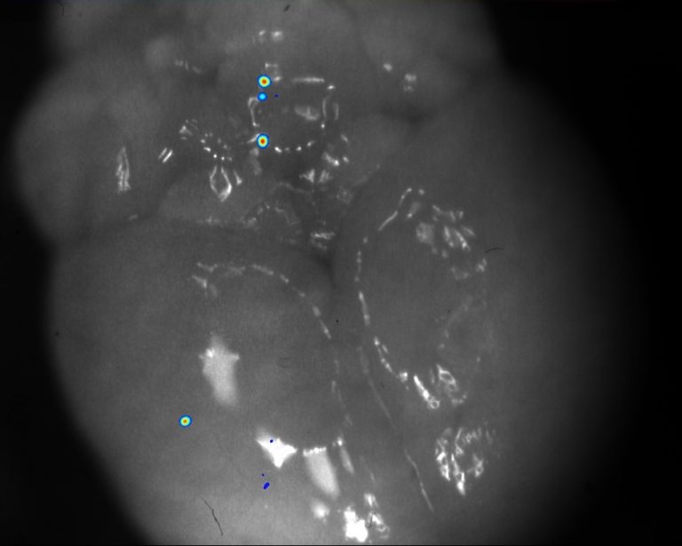
The high sensitivity opens way towards bioluminescence detection to few cells’ detection subcutaneous and few hundreds in deep tissues (livers, lungs, pancreas…)
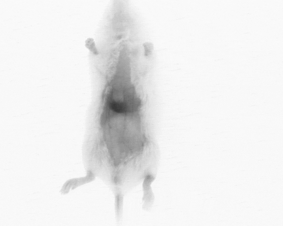
Example of pulmonary metastasis detected in the lungs. Despite the resection, there is a 1,000 factor between the primary tumor and the metastasis.
1- Detect the tiniest biological processes thanks to the unmatched sensitivity of our unique dynamic acquisition technology.
Example of pulmonary metastasis detected in the lungs. Despite the resection, there is a 1,000 factor between the primary tumor and the metastasis.
A single photon detected on the entry face of the intensified camera will generate an intense light spot easily detectable and recognizable by the CCD when intensified.
This technology allows to count individually each photon emitted by the signal center. This method of quantification is called photo counting and leads to an almost perfect linearity as it sums each photon detected.
The photo-counting technology combined with the high-dynamic of the optical chain allow to reach a global linear range over 106 and a linear range within a same image over 104.
The high sensitivity opens way towards bioluminescence detection to few cells’ detection subcutaneous and few hundreds in deep tissues (livers, lungs, pancreas…)
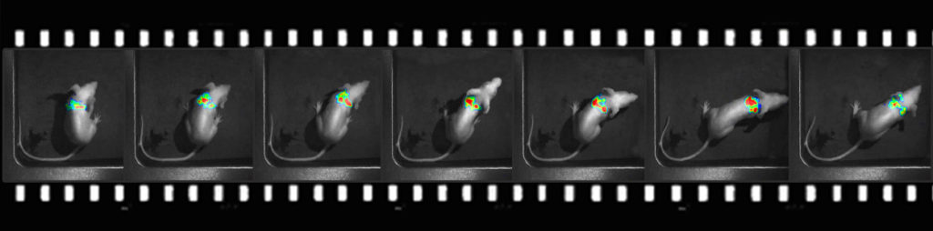
2- Quantify with the greatest accuracy by monitoring the signal kinetics enabled by our real - time acquisition.
Thanks to its high temporal resolution and excellent sensitivity the PhotonIMAGER continuously displays the light distribution from the first seconds of the experiment and can be stopped at any time. The dynamic imaging capability of the PhotonIMAGER system allows the signal kinetics to be followed precisely and independently, even when the signal is weak. It provides precise control over the time-zone selected for the measurement of each ROI, both during and following signal acquisition. The ideal signal measurement window can always be accurately selected for each animal or anatomical region in turn.
Only Real-Time Acquisition can account for the kinetics of the luciferase enzymic reaction and, therefore, allows for accurate quantification. The quantification can then be performed at the optimal time point
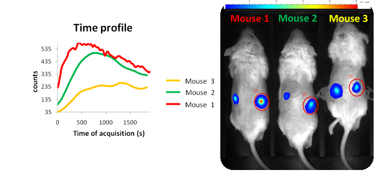
The quantification depends on the biodistribution of the Luciferin (i.e.; anesthesia, food intake, circadian cycle … ). For each acquisition, the signal quantification should be done at the time when the enzyme activity is balanced and the signal intensity issued from it is optimal. It is called the “Plateau”. The kinetics of the signal can vary from one animal to another and is mainly due to marked cells localization – only with real-time acquisition can you choose the temporal windows for quantification on for each emission site.
Although the kinetics are subjected to equivalent protocols and experimental conditions, we can see that the kinetics from emission sites vary and that a reliable quantification requires an individualized plate control.
Picture above shows the signal kinetic from 3 different mice who received a subcutaneous injection of mesenchymal stem cells transfected with the gene expressing the firefly luciferase (Fluc). Each mouse has also been injected in intra-peritoneal with luciferine just before acquisition:
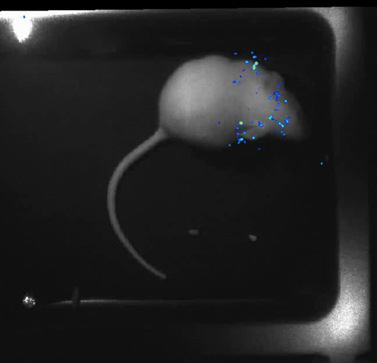
3- Discover unique insights by visualizing moving animals.
With the real-time technology of the PhotonIMAGER, you can even perform experiments with non-anesthetized, freely moving animals, opening previously unimagined new opportunities for research.
Biological phenomena are always dynamic, and so a single observation at a fixed time point often results in an incomplete view of the overall process, which results in misinterpretation of the available data. It results in the loss of information on fast moving dynamic phenomena.
Find out full advantages of imagery of non-anesthetized animals:
Anesthesia is a time-consuming step and reduces the throughput of an imaging system.
Anesthesia can introduce bias by disturbing the physiology and affecting signal expression, so directly affecting the pathology of an animal model. (cf Anesthesia and other considerations for in vivo imaging of small animals, Hildebrandt et al, ILAR 2008).
Imaging an animal in motion is now possible, so monitoring activity in the brain during sleep and wake cycles or observing a bioluminescent signal in a contracting muscle non-invasively in vivo expands research possibilities.
4- A modular system for all applications. Build your own system adapted to your research.
The modular philosophy of the PhotonIMAGER systems allows you to keep up with the latest developments in imaging technologies – existing optional modules and future options can be added to the basic instrument configuration.
Optical imaging has never been easier: change modules quickly, choose your module and acquisition mode on the touchscreen and start acquisition.
MACROLENS
Many real-life applications, such as studying cell invasion and cell migration in Cancer Research or stem cell implantation experiments, require information gathered at different spatial resolutions. The MacroLens module offers a practical solution to this challenge: track cells non invasively at the macroscopic level throughout an entire animal, assess cell growth, spreading or migration at the level of the organ or at a specific anatomic location, focus on smaller groups of cells at the microscopic level.
The MacroLens module was developed by Biospace Lab to open a new window into the world of bioluminescence microscopy and to build the missing link between macroscopic and microscopic for fluorescence and bioluminescence imaging applications.
Macrolens module for high resolution optical imaging down to 2,5µm ex vivo. Zoom in on specific areas of interest and obtain accurate quantification. Correlate the in vivo to ex vivo.
IN ACTIO
The In Actio module simultaneously records both bioluminescence signal and a bright field video of the animal under infra red illumination for co-registration; thereby extending the the imaging capabilities of the Photon Imager from recording dynamic signal from an anesthetized animal, to recording dynamic signal from a free moving animal. The In Actio module offers the following advantages in circumstances where there is no need for multiple views of the animal, as provided by the 3D Module:
Improved physiological relevance: observe physiological phenomena free from any perturbationby anesthesia.
Reduce the time for animal preparation: anesthesia is no longer required and animal stress is reduced
Higher throughput: without anesthesiathe experimental procedure is made faster and several animals can be imaged simultaneously.
4 VIEW – 3D
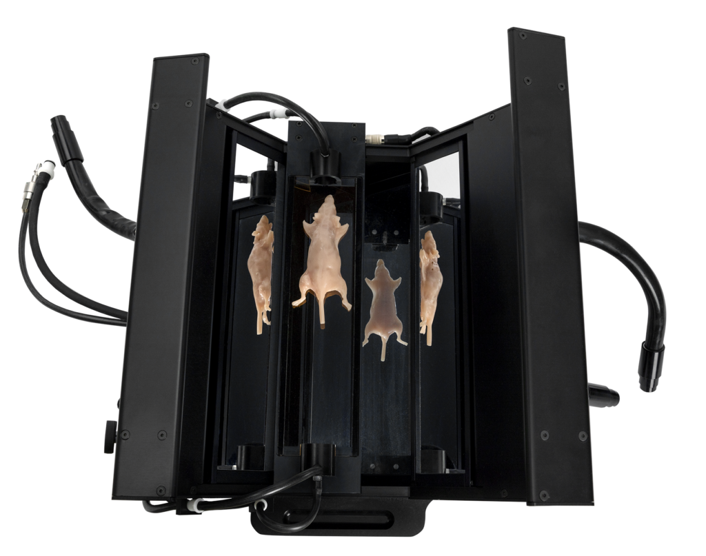
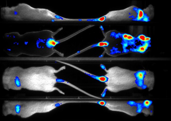
Simultaneous acquisition of the 4 views of the animal The 4 View module helps increasing the analysis accuracy and relevance of your acquisitions. It offers a precise quantification by providing you not only one but several views of your animal (dorsal, ventral, and two lateral views) simultaneously and in real time. The 4-View module is an excellent tool for a better localization of optical signals.
The 4-View – 3D module delivers better localization and quantification of optical signals.
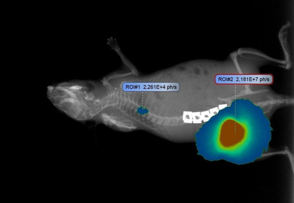
X-RAY
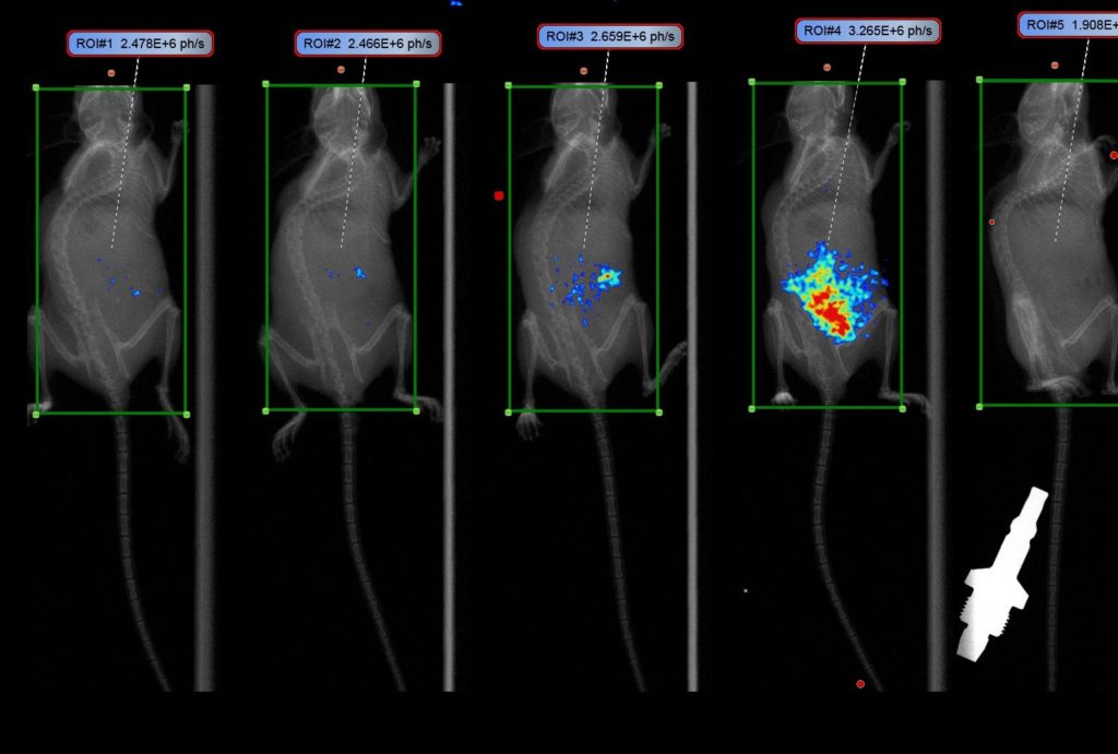
The X-ray module for the PhotonIMAGER Optima provides the capability to overlay x-ray images and optical signals. With this specific, high resolution anatomical information, accurate localization of the optical signals is made easy.
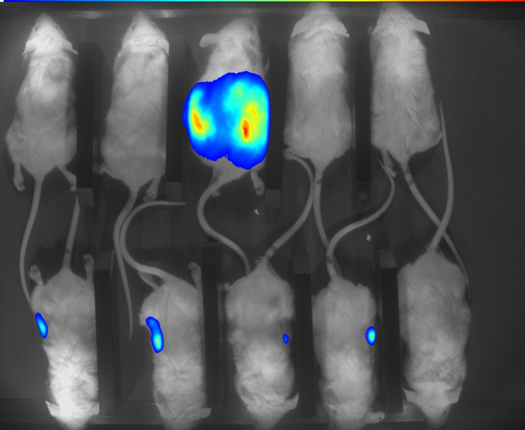
X-ray module provides better anatomical localization of optical signals over other methods. Example demonstrates low dose imaging up to 5 mice.
XRAY
The X-ray module for the PhotonIMAGER Optima provides the capability to overlay x-ray images and optical signals. With this specific, high resolution anatomical information, accurate localization of the optical signals is made easy.
X-ray module provides better anatomical localization of optical signals over other methods. Example demonstrates low dose imaging up to 5 mice.
TOMOFLUO
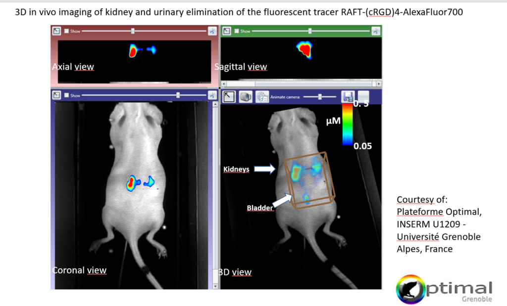
The PhotonIMAGER OPTIMA TomoFluo module was specifically developed for highly precise 3D quantification of fluorescence signals in vivo. It uses fDOT, the next generation of in vivo Fluorescence Tomography algorithms. fDOT (fluorescence Diffusion Optical Tomography) reduces scattering & attenuation artefacts as well as sample boundary problems thanks to its iterative Algebraic Reconstruction Technique (ART). Volumetric Fluoresence data can be overlayed onto a 3D surface reconstruction of the subject or co-registered with CT/PET/SPECT/MRI data.
TomoFluo module enables quantification of deep-seated tissue signals, obtaining accurate pharmacokinetics and longitudinal quantification.
PHOTONIMAGER OPTIMA & RT
PhotonIMAGER Optima
The PhotonIMAGER Optima system is a fully comprehensive solution for advanced in vivo optical imaging applications and is unequalled in its performance.
All application modules are compatible with the PhotonIMAGER Optima.
PhotonIMAGER RT
The PhotonIMAGER RT system is a modular entry level solution for routine in vivo luminescence and fluorescence imaging applications.
Compatible Application Modules: 4 View – 3D, In Actio, Macrolens.
short wave infrared imaging (SWIR / NIR II)
Acquire images with deep penetration, high sensitivity, and high spatial resolution in the short-wave infrared spectrum (SWIR).
No interference from autofluorescence
Minimal light absorption by blood and tissue
Minimized optical scattering
The PhotonIMAGER SWIR is the world’s first integrated commercial solution for preclinical imaging applications in the NIR-II and NIR-III spectrum. Biospace Lab leverages over 30 years of experience in optical imaging to make the SWIR concept an industrial reality.
Biospace Lab is an active member and participant of the NanoTBTech consortium (“Nanoparticles-based 2D thermal bioimaging technologies” FET-Open EU H2020 801305) that develops noninvasive solutions to measure tissue temperature for oncology treatment and diagnosis.
Imaging in the shortwave infrared region (900-1700 nm) improves tissue penetration and spatial resolution. These features enable unprecedented imaging opportunities with direct application to the field of oncology and presents high potential for clinical translation.
Achieve tissue transparency and obtain deep tissue information such as detailed vascular blood flow maps.
Achieving deep photon penetration is one of the main objectives in optical imaging. The NIR II and NIR III regions are characterized by negligible tissue autofluorescence, reduced optical scatter, and lower absorption compared to the near-infrared-I (NIR-I) window (600 to 900 nm). This high spatial resolution capability of the SWIR in combination with high sensitivity resulting from minimal absorption and autofluorescence enables visualization and monitoring of smaller and deeper vessels.
Study rapid biological processes in greater detail than classic imaging modalities.
The PhotonIMAGER SWIR system is a perfect solution to acquire images in real time thanks to its high sensitivity that allows for high frame rates. Contact-free monitoring of respiration and heart rate as well as real time metabolic imaging is possible with unprecedent accuracy.
Customize your own system to respond to your specific applications field, as example the nano-thermometry.
Nano-thermometry is one of the unique and key applications of SWIR imaging, benefiting from the spectral signatures of specialized nanoparticles known as nanothermometers. Nano-thermometry applications include the early detection of cancer, monitoring of enzymatic activity, evaluation of the therapeutic efficiency of drugs and controlling hyperthermia therapy. As nanothermometer probes continue to be developed, temperature sensing rapidly evolves to provide new, indispensable biological information.
Biospace Lab is the only company to offer a nanothermometry module as an integrated option of our PhotonIMAGER SWIR.
Gather more information during your experiments by monitoring the temperature of your region of interest and by using one the 3 following nanothermometry acquisition modes:
Suppression of Intrinsic tissue autofluoresence to obtain superior sensitivity though the fluorescence lifetime imaging mode
Contact us to customize your SWIR instrument for your applications of interest. Choose among different lasers, filters, cameras …
Autoradiography
Acquire ultra-rapid and quantitative images, and capture the small scale biodistributions of low energy radioisotopes as well as multiple isotopes.
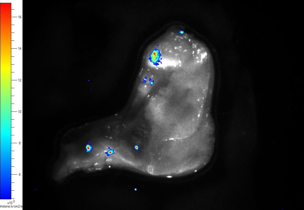
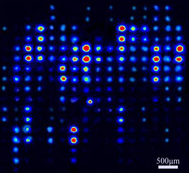
1 - UNIQUE SYSTEM DETECTS AND QUANTIFIES BOTH HIGH AND LOW SAMPLE CONCENTRATIONS IN A SINGLE DATA ACQUISITION WITH BOTH HIGH SENSITIVITY AND RESOLUTION.
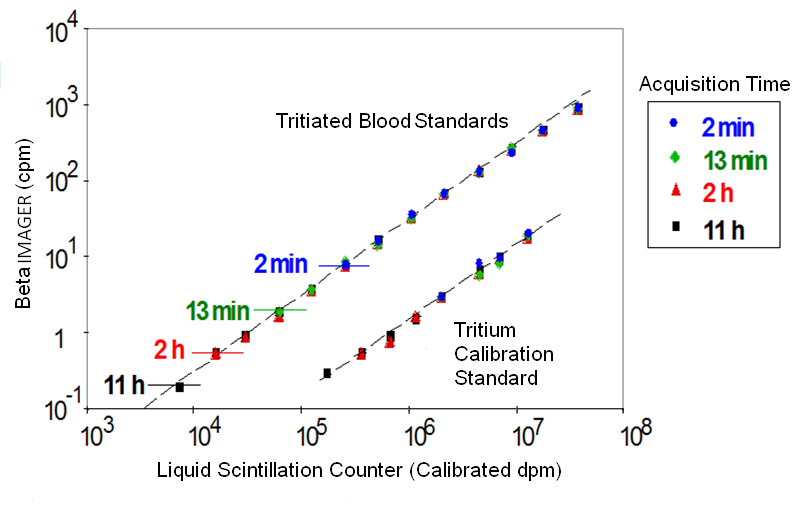
Linear dynamic range of the BetaIMAGER
Courtesy of Dr Alain Schweizer, Novartis
The BetaIMAGER systems count all emitted particles, regardless of their energy. This direct beta counting technology of the BetaIMAGER systems provides unequalled performance: the BetaIMAGER tRACER system is approximately 500X more sensitive and the BetaIMAGER dFINE system about 50X more sensitive to tritium than either X-ray film or Storage Phosphor Screen. The BetaIMAGER tRACER system is unique in its ability to detect tritium levels as low as 0.007 cpm/mm2.
The BetaIMAGER systems provide precise localization and quantification of beta emitting radioligands. The advanced image analysis tools of the M3 vision software allow for direct analysis of digital data and a better than 1:10,000 linear dynamic range ensuring accurate quantification over the widest possible range.
You can detect and quantify both high and low sample concentrations in a single data acquisition.
Biospace Lab developed an exclusive technology for the simultaneous detection and discrimination of multiple radioisotopes.
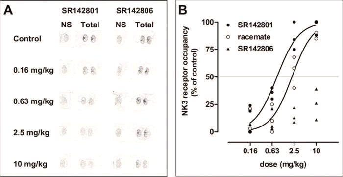
A single sample can be simultaneously labeled with several radioisotopes and the specific contribution from each label can be detected during a single data acquisition. Discrimination can be based on emission energy (2 radioisotopes), or on radioactive half-life (2 isotopes or more). Examples of dual-detection radioisotope pairs include 3H/14C,3H/35S, 3H/32P or 33P and an example of a multi-label combination would be 99mTc/ 111In/18F.
This unique radioisotope discrimination feature allows the development of more rapid and accurate protocols for recording the distribution of several molecules simultaneously in a single specimen.
You can determine co-localization of different receptors, or receptors and ligands; Map complex functional activity with pathology; or Compare specific gene expression with a reference gene.
1 - UNIQUE SYSTEM DETECTS AND QUANTIFIES BOTH HIGH AND LOW SAMPLE CONCENTRATIONS IN A SINGLE DATA ACQUISITION WITH BOTH HIGH SENSITIVITY AND RESOLUTION.
The BetaIMAGER™ systems count all emitted particles, regardless of their energy. This direct beta counting technology of the BetaIMAGER systems provides unequalled performance:
the BetaIMAGER tRACER system is approximately 500X more sensitive and the BetaIMAGER dFINE system about 50X more sensitive to tritium than either X-ray film or Storage Phosphor Screen. The BetaIMAGER™ tRACER system is unique in its ability to detect tritium levels as low as 0.007 cpm/mm2.
The BetaIMAGER systems provide precise localization and quantification of beta emitting radioligands. The advanced image analysis tools of the M3 vision software allow for direct analysis of digital data and a better than 1:10,000 linear dynamic range ensuring accurate quantification over the widest possible range.
Linear dynamic range of the BetaIMAGER
Courtesy of Dr Alain Schweizer, Novartis
You can detect and quantify both high and low sample concentrations in a single data acquisition.
Biospace Lab developed an exclusive technology for the simultaneous detection and discrimination of multiple radioisotopes
A single sample can be simultaneously labeled with several radioisotopes and the specific contribution from each label can be detected during a single data acquisition. Discrimination can be based on emission energy (2 radioisotopes), or on radioactive half-life (2 isotopes or more). Examples of dual-detection radioisotope pairs include 3H/14C,3H/35S, 3H/32P or 33P and an example of a multi-label combination would be 99mTc/ 111In/18F
This unique radioisotope discrimination feature allows the development of more rapid and accurate protocols for recording the distribution of several molecules simultaneously in a single specimen.
You can determine co-localization of different receptors, or receptors and ligands; Map complex functional activity with pathology; or Compare specific gene expression with a reference gene.
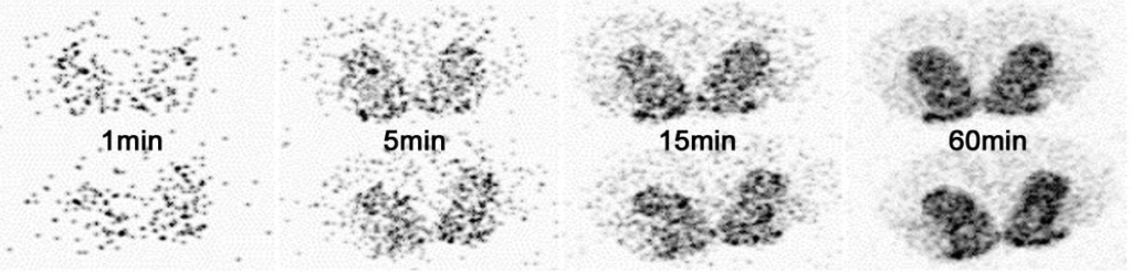
2- SAVE hours or days by acquiring images in real time thanks to our real time detection technology.
The exclusive real-time detection technology used in both systems displays and accumulates counts in real-time:
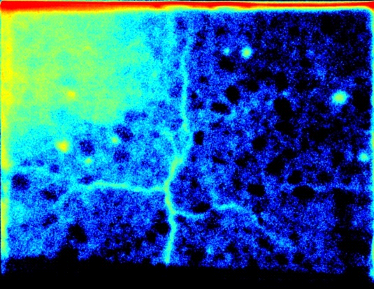
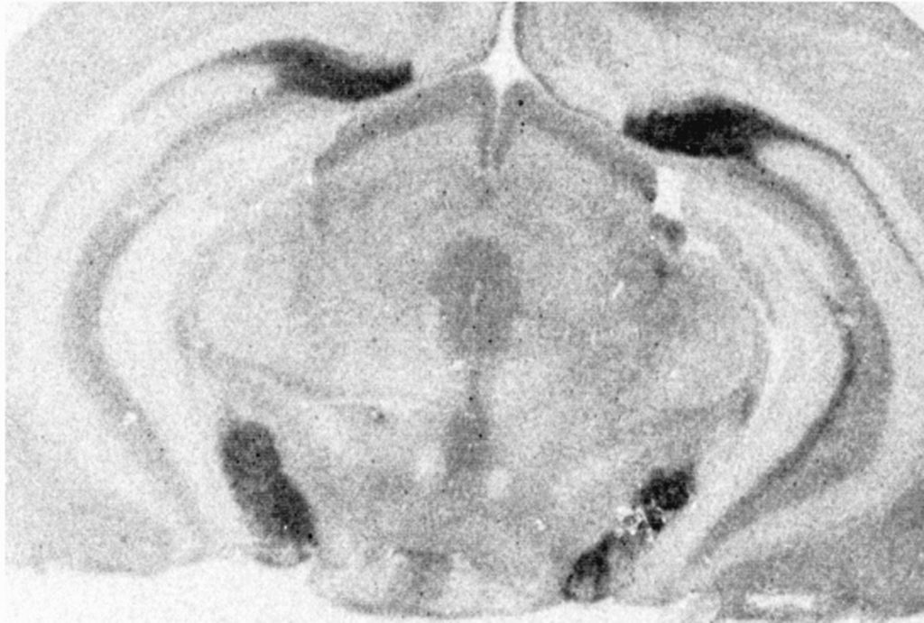
Real-time autoradiography: distribution of D1 receptors in rat brain striatum following tritiated ligand binding
X-Ray film or Storage Phosphor screen based autoradiography requires a two-step blind acquisition process: expose and then scan
The real-time acquisition allows the instantaneous display of the image recorded by the camera and to follow the progression of the reconstitution of the finale image. The image display is refreshed every 30 seconds allowing the user to evaluate it during the acquisition.
The unmatched sensitivity reduces your data acquisition time from weeks to hours.
You can see data accumulating in real-time and obtain the first results in minutes.
3- NO LOST DATA OR EXPERIMENTS CAUSED BY SATURATION.
Disintegrations are detected in real-time and the data display is continuously updated during acquisition. Under or over exposure of the sample are eliminated.
Longer acquisitions deliver higher image dynamics. The absence of limit on the duration of the acquisition allows the detection of weak signals and the improvement of resolution.
4- Use minimal amounts of radiolabeled compounds, reduce radioactive waste and minimize experimental costs as you can detect ultra-low level of bounds radiolabeled ligand.
Thanks to their high sensitivity, BetaIMAGER systems can detect ultra-low levels of radiolabeled ligands in the sample. You can use minimal amounts of radiolabeled compounds and reduce radioactive waste as well as minimize your experimental costs.
Reduce the number of experiments thanks to the reproducibility of the system.
Non-destructive imaging and measurement: specimens are never damaged or destroyed during the process.
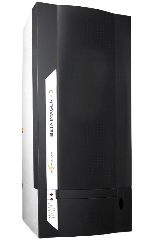
BETAIMAGER SYSTEMS
Whether high speed or high-resolution, Biospace Lab has the ideal solution for your autoradiography application.
BETAIMAGER TRACER
The BetaIMAGER™ tRACER is the system of choice for applications requiring the highest sensitivity and either high throughput, or larger sample sizes. With the BetaIMAGER tRACER, autoradiography becomes a fast and easy routine.
Tritium is a reliable beta emitter for measurement of biological samples. The big problem is that detection is slow. Traditional film and phosphor imaging methods can take weeks to complete. But now, there’s a better way to meet the high throughput needs of your lab: The BetaIMAGER tRACER system cuts your tritium imaging time from weeks to hours. Exceptional sensitivity can be obtained even with ultra-low energy tritium emissions: the tRACER system detects tritium levels as low as 0,007 cpm/mm2
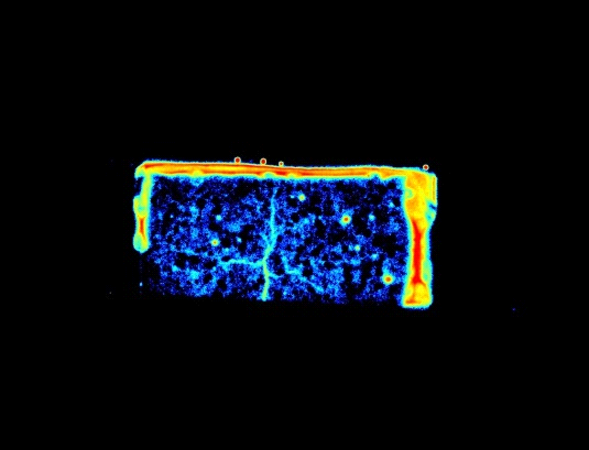
With a 200 x 250mm maximum field of view, the BetaIMAGER can accommodate up to 15 microscope slides, several larger tissue sections, or TLC plates for simultaneous image acquisition.
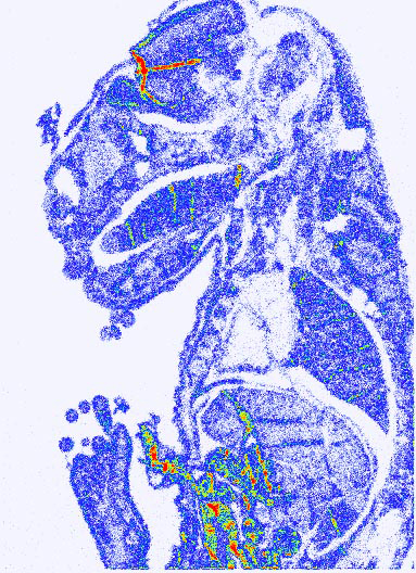
Sample activity was calculated using the acquisition shown in the figure on the left (exhibits the entire sample): 7,5.103 hpm (hits per minute) which translates to 6,82 nCi.
Tritiated sample. 5h acquisition. Wider visual range than the sample
The BetaIMAGER™ tRACER is the system of choice for applications requiring the highest sensitivity and either high throughput, or larger sample sizes. With the BetaIMAGER tRACER, autoradiography becomes a fast and easy routine.
Tritium is a reliable beta emitter for measurement of biological samples. The big problem is that detection is slow. Traditional film and phosphor imaging methods can take weeks to complete. But now, there’s a better way to meet the high throughput needs of your lab: The BetaIMAGER tRACER system cuts your tritium imaging time from weeks to hours. Exceptional sensitivity can be obtained even with ultra-low energy tritium emissions: the tRACER system detects tritium levels as low as 0,007 cpm/mm2.
With a 200 x 250mm maximum field of view, the Beta Imager can accommodate up to 15 microscope slides, several larger tissue sections, or TLC plates for simultaneous image acquisition.
Tritiated sample. 5h acquisition. Wider visual range than the sample
Sample activity was calculated using the acquisition shown in the figure on the left (exhibits the entire sample): 7,5.103 hpm (hits per minute) which translates to 6,82 nCi.
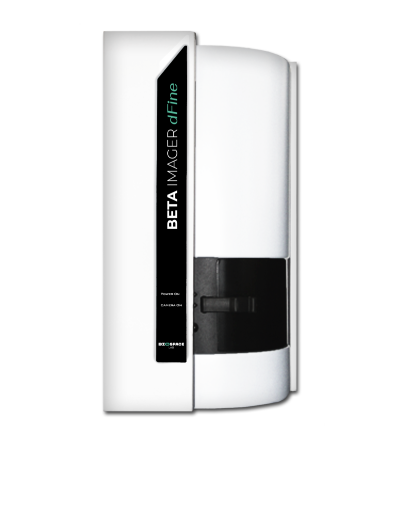
BETAIMAGER DFINE
The ONLY high-resolution digital autoradiography solution.
The BetaIMAGER™ DFINE is the system of choice for applications involving smaller tissue sections and where the best possible spatial resolution is required. Four samples, up to 24 x 32mm each in size, can be loaded onto the automated stage for sequential image acquisition. Spatial resolution as high as 10μm can be achieved with tritium. The DFINE is the perfect ex-vivo complement to PET and SPECT molecular imaging.
The BetaIMAGER DFINE is 50 times faster than film with tritium-labeled samples, allowing precise quantitation. The DEFINE boasts a 10μm resolution for tritiated samples and is unsurpassed by any other non-film technique. Further, DFINE has no complex techniques or procedures to learn.
2 - The BetaIMAGER™ DFINE
The ONLY high-resolution digital autoradiography solution.
The BetaIMAGER™ DFINE is the system of choice for applications involving smaller tissue sections and where the best possible spatial resolution is required. Four samples, up to 24 x 32mm each in size, can be loaded onto the automated stage for sequential image acquisition. Spatial resolution as high as 10μm can be achieved with tritium. The DFINE is the perfect ex-vivo complement to PET and SPECT molecular imaging.
The BetaIMAGER DFINE is 50 times faster than film with tritium-labeled samples, allowing precise quantitation. The DEFINE boasts a 10μm resolution for tritiated samples and is unsurpassed by any other non-film technique. Further, DFINE has no complex techniques or procedures to learn.
Software
The IMAGER systems are driven by the PhotoAcquisition and BetaAcquisition software which seamlessly control the entire system and its modules, and manage the imaging experiments.
The user-friendly interface of the software makes the use of the instrument simple and pleasant.
Simplicity and user-friendly Acquisition software
Immediate visualization from the beginning of acquisition is one of the hallmarks making real-time imagery so practical. You can see the first signals after just a few seconds and can quickly stop the acquisition or extend it knowing that the signal will continue to accumulate until the desired endpoint.
Real-time feedback allows for optimized and automated control of some acquisition parameters such as the opening of the lens during the acquisition depending on the signal progress. These automatic time optimization capabilities simplify the use without blind configuration of the lens opening, integration time or binning level which must often be determined experimentally on the cooled CCD camera system.
Simplicity of the analysis software
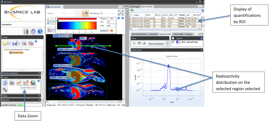
M3Vision analysis software allows users to visualize 2D and 3D images, freely adjust the signal display threshold, and perform quantifications based on desired ROIs such as geometrical forms or signal intensity thresholds.
The measurements made on the ROIs will allow a quantification of the total value of the signal, its top intensity, its lowest and its average value.
M3Vision makes time analysis possible, allowing quantifications at multiple ROIs at the plateau time. For high speed studies, an automatic selection tool of the temporal window to the signal plateau allows the rapid analysis of each ROI.
Export all data, measurements and time profiles to Excel in a single click! For the longitudinal studies, all acquisitions linked to a study can be exported in a single Excel folder that includes one tab per acquisition as well as a summary table of all the quantifications.
Software analysis tools include numerous functions to allow precise and automatic quantification:
The magic wand tool allows you to automatically draw ROIs determined by signal intensity thresholds. These ROIs can then be created and analyzed automatically by the software in a fraction of a second.
Automatic temporal analysis enables automated, streamlined and rapid quantification on each ROI in just a few seconds.
Traceability
When an acquisition is saved, ALL acquisition settings (including lens opening, plate position, acquisition time, grid speed, date and time) are also saved. User-generated comments can also be saved such as observations about the animals being imaged, and notable events that occurred before or during acquisition. This information is linked to the acquisition folder and is accessible through the analysis software.